
重磅活動 冷凍電鏡樣品制備前沿技術演示及研討會

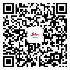
掃描二維碼
報名參與活動
特別說明:
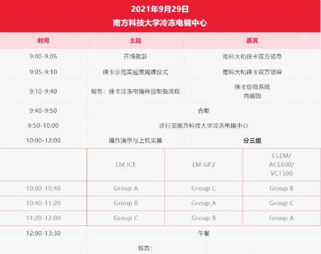
1. 本次活動全天直播
2. 鑒于疫情管控因素,推薦廣大校外學員線上參加研討會
3. 設置討論與交流環節,歡迎廣大學員積極提問
4. 本次研討會免費
活動主辦方:
特邀報告人

劉錚
南方科技大學冷凍電鏡中心 教授
1997年取得清華大學生物物理專業博士學位,2005年在美國Wadsworth Center任研究員,2015年兼同濟大學醫學院泛血管研究所副所長,心肺血管研究所副所長,博士生導師。2020年初入職南方科技大學冷凍電鏡中心。
主要通過冷凍電子顯微學、生物化學、細胞生物學等多種技術手段研究細胞器、膜蛋白等生物大分子的工作機制。先后在國內外重要學術期刊發表相關研究成果,其中SCI收錄40余篇。

祝建
同濟大學 教授
- 1982年畢業于寧夏大學農學院,留校任教
- 1992—1995年蘇黎世瑞士聯邦理工學院(ETH)細胞生物所電鏡技術實驗室,中瑞聯合培養博士
- 1996—2000年上海鐵道大學醫學院
- 2000—2018年同濟大學生命科學與技術學院,生物電鏡技術實驗室,教授,博導

ROLAND A. FLECK
Professor of Ultrastructural Imaging, Royal Society Industry Fellow and Director of the Centre for Ultrastructural Imaging at King's College London, United Kingdom.
He is also a visiting Professor of the Faculty of Health and Medical Sciences, University of Copenhagen and Professor of the UNESCO Chair in Cryobiology, National Academy of Sciences of Ukraine, Institute for Problems of Cryobiology, Kharkiv, Ukraine. He joined King’s College London in 2013 from the National Institute for Biological Standards and Control (NIBSC), where he was head of Biological Imaging and Assay Development. At NIBSC he developed advanced imaging techniques for the control and standardisation of biological medicines and had research interests in developing differentiation protocols for myeloid leukemic and human embryonic stem cell lines as substrates for functional biological assays. He has extensive specialist knowledge of freeze fracture/freeze etch preparation of tissues and wider cryo-microscopic techniques.
As academic director of the Centre for Ultrastructural Imaging he collaborates widely with colleagues in neuroscience and parasitology and promotes advanced three dimensional studies of cells and tissues using both room temperature and cryo electron microscopy techniques. He has extensive experience and knowledge of low temperature biology and cryopreservation having researched how cells and tissues both avoid and are damaged by chilling and freezing events. His current research interests focus on developing tools and protocols for enhancing the preservation of tissues for characterisation by electron microscopy as a capacity to enhance wider scientific collaborations.
Areas of expertise: Advanced electron microscopy techniques, cryo electron microscopy preparation techniques and electron tomography, application of serial block face and focused ion beam for the life sciences.

Jan De Bock
Jan studied biology and did his PhD in the field of olfaction, characterizing olfactory neurons in their response to odorants.
Jan has worked as a microscopy expert in different roles since 2003. He joined Leica Microsystems in 2011 as a product specialist for confocal microscopy. In 2017, he became member of the newly formed Workflow and Application Team responsible for correlative workflows, in particular involving sample preparation and imaging under cryogenic conditions.
Abstract
Cryo Electron Microscopy workflows are a state-of-the-art tool to investigate proteins in the cellular context with subnanometer resolution. To succeed in this, light microscopes performing under cryogenic conditions are essential for an early quality check and to identify target structures for the subsequent EM analysis.
In this webinar we show Leica Microsystem´s cryo microscope solutions for the assessement of the sample quality, a safe sample transfer and super resolution imaging under cryogenic conditions as a basis for precise targeting.
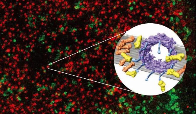
冷凍電子斷層圖像。與核孔復合體相連的蛋白酶體(紫色)。由分子結構系Ben Engel博士提供。生物學,生物化學MPI,馬丁斯利德,德國
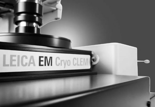
Leica EM Cryo CLEM
細胞內大分子原位結構研究
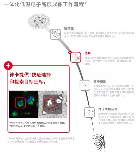
徠卡顯微系統可為不同品牌SEM-FIB雙束顯微鏡定制提供完全集成的低溫電子斷層成像工作流程,以滿足研究需要。
了解更多:徠卡顯微

- 喜報:普瑞邦中標中儲糧,攜手守護大國糧倉
- 廣州市工信局蒞臨走訪祝賀艾貝泰入圍揭榜掛帥名單
- 喜訊:艾普拜榮獲年度省級“專精特新”中小企業認定
- SPT Labtech 與水木未來共建冷凍制樣創新實驗室
- 明美MIX60+MS23成像設備助力廈門大學微流控成像升級
- 易科泰葉綠素熒光成像系統落地上海頂尖科研機構
- 理加聯合出席云南省自然生態監測網絡工作培訓會
- 博格隆榮獲十四五中國醫藥產業優質供應商100稱號
- 沃德精準攜多款重磅智能監測設備亮相深圳環博會
- 易科泰植物逆境模擬與生長監測系統落戶寧夏農科院
- 上海金鵬助力首屆蛋白質純化和分析技能賽項圓滿成功
- 喜訊:固康生物成為意大利DiaMetra公司中國經銷商
- 上海生物芯片推出SMI WTX與空間蛋白聯合檢測方案
- 明美顯微鏡助力中科院深圳先進院合成所活細胞研究
- 隱智科儀中標合肥師范學院實驗室搬遷服務項目
- 明美光電獲授國家級“專精特新”小巨人牌匾
- 明美顯微鏡助力博物館重現千年微觀之美
- 奧龍集團參加遼寧省工業互聯網實驗室群成立大會
- 徠卡生命科學部積極響應,助力國家以舊換新政策
- 明美與您相約國際檢驗醫學暨輸血儀器試劑博覽會
- 直播預告:FALCON-快速熒光壽命成像應用進展
- 再創里程碑,徠卡顯微系統廣州客戶體驗中心正式啟動
- 2023年徠卡工業顯微鏡用戶作品有獎征集大賽通知
- 北京佰司特中標大連理工超高速視頻級原子力顯微鏡
- 明美LED熒光光源助力深圳婦幼保健院免疫熒光觀察
- 明美倒置熒光顯微鏡走進天津血液研究所
- 明美推出新品活細胞成像儀MCS11/MCS21
- 明美推出新品大體成像儀,讓標本拍攝更簡單
- 蔡司全新Mineralogic 3D開啟三維礦物分析新紀元
- 重磅活動 冷凍電鏡樣品制備前沿技術演示及研討會